INSTITUTE OF CYTOLOGY OF THE RUSSIAN ACADEMY OF SCIENCES

Laboratory of Cell Morphology

    Head of the Laboratory


    Dmitry S. BOGOLYUBOV

    D.Sci.

    Phone: +7 (812) 297-18-36
    e-mail: dbogol@mail.ru


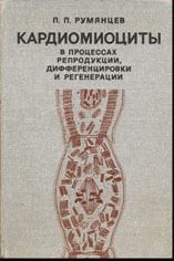
The Laboratory of Cell Morphology exists since the Institute of Cytology was established. The Laboratory was found by the eminent cytogenetists and zoologist I.I. Sokolov. He was the head of the Laboratory since 1957 until 1959. Professor Sokolov placed high emphasis on the studies of gametogenesis, particularly oogenesis. Then, the Laboratory was headed by Professor L.N. Zhinkin (since 1959 until 1971) who extended the frame of studies to histogenesis, mitotic cycle, differentiation and regeneraton of the somatic cells (mainly muscle cells) and generally introduced the autoradiography as a new method. Professor P.P. Rumyantsev, a vice academician of Soviet Academy of Sciences, was the head of the Laboratory since 1971 until 1988. He established a new line of investigation: the study of cardiomyocytes. Together with his apprentices, he brilliantly developed this line being prominent world-wide specialist in this field.

Since 1988 until 2012, the Laboratory was headed by Professor V.N. Parfenov. He had begun his scientific career here as a post-graduate student and then got the position of Head of the Laboratory and the Director of the Institute. Professor Parfenov gave a new impulse for studies of the organization and function of oocyte nuclear structures keeping at the same time all previous lines of studies. He extended the range of objects and contributed some pioneering methods including immunocytochemistry on light and electron microscopic (immunogold) levels.

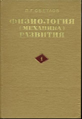
In 1957-1968, a noted cytologist, future head of the Department of Histology and Cytology at the Saint-Petersburg University, Professor A.A. Zavarzin, Jr. worked at the Laboratory. In 1966-1974, the first-rate embryologist, a vice academician of Soviet Academy of Medical Sciences, Professor P.G. Svetlov also worked there.

The main part of studies is being carried out in the frame of the interlaboratory government topic "Molecular structure, composition and dynamics of chromosomes and the extrachromosomal nucleoplasmic domains of different cell types in phylogenetically distant organisms" (No. 01201351105).

In 1988, a series of publications by E.V. Raikova on the cytology of the unique intracellular parasite of acipenserid oocytes, Polypodium hydriforme (Cnidaria), and the cytology of the oogenesis of acipenseriform fishes was awarded by the Alexander Kowalevsky Medal from the Presidium of Soviet Academy of Sciences.

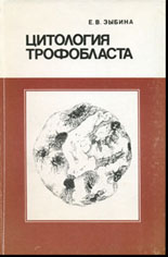
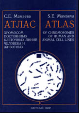
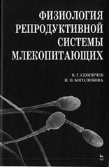
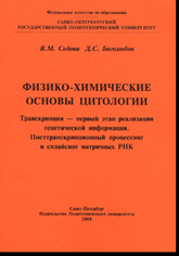
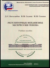
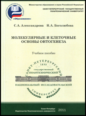
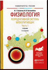
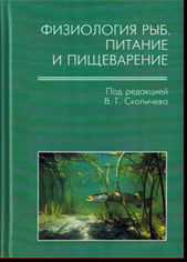
In different years, the collaborators of the Laboratory have written and published some monographs, textbooks and manuals on the cytology:


At the present time, the collaborators of the Laboratory give courses of lectures at Saint-Petersburg State University, Saint-Petersburg Polytechnic University, Central Lecture Center, medical institutions and universities of Saint-Petersburg and take part in writing of textbooks and manuals for students.

The work was repeatedly supported by grants of Fogarty Foundation, Russian Fund for Basic Research, Soros Foundation, National Science Support Foundation, Government of Saint-Petersburg, Presidium of Russian Academy of Sciences, and the grant program "Leading Scientific Schools of Russian Federation".


    LABORATORY STAFF

    Chief Research Associate

    Dmitry S. Bogolyubov, Ph.D., D. Sci.

    Principal Research Associates
    Marina G. Martynova, Ph.D., D. Sci.

    Senior Research Associates
    Natalia A. Bogoliubova, Ph.D.
    Irina O. Bogolyubova, Ph.D.
    Olga A. Bystrova, Ph.D.
    Sergej Yu. Demin, Ph.D.
    Galina N. Pochukalina, Ph.D.
    Tatiana K. Yakovleva, Ph.D.

    Research Associates
    Marina I. Krylova, Ph.D.
    Irina S. Stepanova, Ph.D.

    Junior research Associates
    Maxim I. Sulatsky
    Viktoria I. Turilova
    Natalia M. Yartseva

    Senior Laboratory Assistants
    Olga I. Emelyanova
    Ksenia V. Tyutina

    Technician
    Yekaterina V. Surko


Lines of investigations

The investigations at the Laboratory are carried out on wide-ranged objects using complex approaches including light and electron microscopy, cyto- and histochemistry, light and electron autoradiography, cytophotometry, indirect light and electron (immunogold) microscopy, confocal laser scanning microscopy, hybridization in situ, microinjections, protein electrophoresis, Western blotting, etc.

At the present time, the studies are carried out on the following lines:

  • I. FUNCTIONAL MORPHOLOGY OF NUCLEAR STRUCTURES IN OOGENESIS
  • II. FUNCTIONAL MORPHOLOGY OF THE NEUROENDOCRINE SYSTEM
  • III. CYTOGENETICS
  • IV. PROBLEMS OF EARLY EMBRYOGENESIS OF MAMMALS
  • PROBLEMS OF TROPHOBLAST REPRODUCTION (until 2018)
  • CYTOLOGY OF INTRACELLULAR PARASITISM AND PARASITE-HOST RELATIONS (until 2018)

  • I. FUNCTIONAL MORPHOLOGY OF NUCLEAR STRUCTURES IN OOGENESIS

    The Head: D.S. Bogolyubov, Ph.D, D.Sci.

    Complex studies of the organization, dynamics, molecular composition and functions of chromosomal and nucleolar apparatus as well as extrachromosomal nuclear domains of oocytes are carried out on broad-ranged objects including some platyhelmints, mollusks, different insects, amphibians, and mammals. The main attention is paid to the revealing of peculiarities and the significance of the relations between Cajal bodies, nucleoli and interchromatin granule clusters (nuclear speckles) at active stages of oogenesis and under physiologically determined inactivation of oocyte nuclei towards the end of oogenesis during karyosphere (karyosome) formation.

    Main publications (1993-2018)

    Gruzova M.N., Parfenov V.N. (1993) Karyosphere in oogenesis and intranuclear morphogenesis. Int. Rev. Cytol. 144: 1-52.

    Wu Z., Murphy C., Wu C.-H.H., Tsvetkov A., Gall J.G. (1993) Snurposomes and coiled bodies. Cold Spring Harb. Symp. Quant. Biol. 58: 747-754.

    Gall J.G., Tsvetkov A., Wu Z, Murphy C. (1995) Is the sphere organelle/coiled body a universal nuclear component? Dev. Genet. 16: 25-35.

    Parfenov V.N., Davis D.S., Pochukalina G.N., Sample C.E., Bugaeva E.A., Murti K.G. (1995) Nuclear actin filaments and their topological changes in frog oocytes. Exp. Cell Res. 217: 385-394.

    Parfenov V.N., Davis D.S., Pochukalina G.N., Sample C.E., Murti K.G. (1996) Nuclear bodies of stage 6 oocytes of Rana temporaria contain nucleolar and coiled body proteins. Exp. Cell Res. 228: 229-236.

    Tsvetkov A., Alexandrova O., Bogolyubov D., Gruzova M. (1997) Nuclear bodies from the cricket and mealworm oocytes contain splicing factors of pre-mRNA. Eur. J. Entomol. 94: 393-407.

    Parfenov V.N., Davis D.S., Pochukalina G.N., Kostyuchek D.F., Murti K.G. (1998) Dynamics of distribution of splicing components relative to the transcriptional state of human oocytes from antral follicles. J. Cell. Biochem. 69: 72-80.

    Bogolyubov D., Alexandrova O., Tsvetkov A., Parfenov V. (2000) An immunoelectron study of karyosphere and nuclear bodies in oocytes of mealworm beetle, Tenebrio molitor (Coleoptera: Polyphaga). Chromosoma. 109: 415-425.

    Parfenov V.N., Davis D.S., Pochukalina G.N., Kostyuchek D. Murti K.G. (2000) Nuclear distribution of RNA polymerase II in human oocytes from antral follicles: dynamics relative to the transcriptional state and association with splicing factors. J. Cell. Biochem. 77: 654?665.

    Bogolyubov D., Parfenov V. (2001) Immunogold localization of RNA polymerase II and pre-mRNA splicing factors in Tenebrio molitor oocyte nuclei with special emphasis on karyosphere development. Tiss. Cell. 33: 549-561.

    Parfenova H., Parfenov V.N., Shlopov B.V., Levine V., Falkos S., Pourcyrous M., Leffler C.W. (2001) Dynamics of nuclear localization sites for COX-2 in vascular endothelial cells. Am. J. Physiol. Cell. Physiol. 281: C166-C178.

    Batalova F., Parfenov V. (2003) Immunomorphological localization of Vasa protein and pre-mRNA splicing factors in Panorpa communis trophocytes and oocytes. Cell Biol. Int. 27: 795-807.

    Parfenov V.N., Pochukalina G.N., Davis D.S., Reinbold R., Scholer H.R., Murti K.G. (2003) Nuclear distribution of Oct-4 transcription factor in transcriptionally active and inactive mouse oocytes and its relation to RNA polymerase II and splicing factors. J. Cell. Biochem. 89: 720-732.

    Bogolyubov D., Parfenov V. (2004) Do nuclear bodies in oocytes of Tenebrio molitor (Coleoptera: Polyphaga, Tenebrionidae) contain two forms of RNA polymerase II? Tiss. Cell. 36: 13-17.

    Batalova F.M., Stepanova I.S., Skovorodkin I.N., Bogolyubov D.S., Parfenov V.N. (2005) Identification and dynamics of Cajal bodies in relation to karyosphere formation in scorpionfly oocytes. Chromosoma. 113: 428-439.

    Parfenova H., Parfenov V., Leffler C. (2005) Nuclear functions of cyclooxygenase in cerebral vascular endothelial cells. In: Biochemistry of Lipids in Vascular Functions. M. Alberghina, ed. pp. 63-83.

    Stepanova I.S., Bogolyubov D.S., Skovorodkin I.N., Parfenov V.N. (2007) Cajal bodies and interchromatin granule clusters in cricket oocytes: composition, dynamics and interactions. Cell Biol. Int. 31: 203-214.

    Bogolyubov D. (2007) Localization of RNA transcription sites in insect oocytes using microinjections of 5-bromouridine 5 -triphosphate. Folia Histochem. Cytobiol. 45: 129-134.

    Bogolyubov D.S., Batalova F.M., Ogorzalek A. (2007) Localization of interchromatin granule cluster and Cajal body components in oocyte nuclear bodies of the hemipterans. Tissue Cell. 39: 353-364.

    Bogolyubov D., Stepanova I. (2007) Interchromatin granule clusters in vitellogenic oocytes of the fleshfly, Sarcophaga sp. Folia Histochem. Cytobiol. 45: 401-403.

    Dolnik A.V., Pochukalina G.N., Parfenov V.N., Karpushev A.V., Podgornaya O.I., Voronin A.P. (2007) Dynamics of satellite binding protein CENP-B and telomere binding protein TRF2/MTBP in the nuclei of mouse spermatogenic line. Cell Biol. Int. 31: 316-329.

    Tolkunova E., Malashicheva A., Parfenov V.N., Sustmann C., Grosschedl R., Tomilin A. (2007) PIAS proteins as repressors of Oct4 function. J. Mol. Biol. 374: 1200-1212.

    Bogolyubov D., Parfenov V. (2008) Structure of the insect oocyte nucleus with special reference to interchromatin granule clusters and Cajal bodies. Int. Rev. Cell Mol. Biol. 269: 59-110.

    Bogolyubov D., Stepanova I., Parfenov V. (2009) Universal nuclear domains of somatic and germ cells: some lessons from oocyte interchromatin granule cluster and Cajal body structure and molecular composition. BioEssays. 31 (4): 400-409.

    Batalova F., Parfenov V. (2009) Factors related to RNA polymerase II transcription are localized in interchromatin granule clusters of Panorpa communis oocytes. Folia Histochem. Cytobiol. 47 (1): 123-126.

    Batalova F.M., Bogolyubov D.S., Parfenov V.N. (2010) Interchromatin granule clusters of the scorpionfly oocytes contain poly(A)+ RNA, heterogeneous ribonucleoproteins A/B, and mRNA export factor NXF1. Cell Biol. Int. 34 (12): 1163-1170.

    Bogolyubov D.S., Kiselyov A.M., Shabelnikov S.V., Parfenov V.N. (2012) Polyadenylated RNA and mRNA export factors in extrachromosomal nuclear domains of vitellogenic oocytes in the yellow mealworm Tenebrio molitor. Cell Tissue Biol. 6: 412-422.

    Pochukalina G.N., Parfenov V.N. (2012) Localization of actin and mRNA export factors in the nucleus of murine preovulatory oocytes. Cell Tissue Biol. 6: 423-434.

    Bogolyubova I.O., Bogolyubov D.S. (2013). Oocyte nuclear structure during mammalian oogenesis. In: Recent Advances in Germ Cells Research. (Perrotte A., Ed.). Nova Biomedical. pp. 105-132.

    Bogolyubov D.S., Batalova F.M., Kiselyov A.M., Stepanova I.S. (2013). Nuclear structures in Tribolium castaneum oocytes. Cell Biol. Int. 37: 1061-1079.

    Pochukalina G.N., Stepanova I.S. 2014. Interchromatin granule clusters of mouse preovulatory oocytes are enriched with some components of mRNA export machinery. Annu. Res. Rev. Biol. 4: 79-92.

    Ilicheva N.V., Kiryushina D.Y., Baskakov A.V., Podgornaya O.I., Pochukalina G.N. 2016. The karyosphere capsule in oocytes of hibernating frogs Rana temporaria contains actin, lamins and SnPNP. Cell Tiss. Biol. 10: 422–429.

    Pochukalina G.N., Ilicheva N.V., Podgornaya O.I., Voronin A.P. 2016. Nucleolus-like body of mouse oocytes contains lamin A and B and TRF2 but not actin and topo II. Mol. Cytogen. 9: 50.

    Stepanova I.S., Bogolyubov D.S. 2017. Localization of the chromatin-remodeling protein ATRX in the oocyte nucleus of some insects. Cell Tiss. Biol. 11: 416–425.

    Kiselev A.M., Stepanova I.S., Adonin L.S., Batalova F.M., Parfenov V.N., Bogolyubov D.S., Podgornaya O.I. 2017. The exon junction complex factor Y14 is dynamic in the nucleus of the beetle Tribolium castaneum during late oogenesis. Mol. Cytogenet. 10 : 41.

    Bogolyubov D. 2018. Karyosphere (karyosome): A peculiar structure of the oocyte nucleus. Int. Rev. Cell Mol. Biol. 337: 1–48.

    Ilicheva N., Podgornaya O., Bogolyubov D., Pochukalina G. 2018. The karyosphere capsule in Rana temporaria oocytes contains structural and DNA-binding proteins. Nucleus. 9 (1): 516–529.


    * The names of the laboratory staff are in bold.                              Updated: January 2018
    II. FUNCTIONAL MORPHOLOGY OF THE NEUROENDOCRINE SYSTEM

    The Head: - M.G. Martynova, Ph.D., D.Sci.


    The mainstream of the research is connected with the studying of vertebrate mast cells and their functional analogs in invertebrates. The interactions of these cells with the nerve and circulatory systems are being studied. Furthermore, staff of the group also continues traditional studies in the field of cardiomyogenesis, and also participates in joint studies with the staff of other laboratories of the Institute and medical organizations of St. Petersburg in the field of studying of human stem cells and development of nanomedicine techniques for brain-tumor therapy.

    Main publications (1990-2018)

    Rumyantsev P.P., Erokhina I.L., Antipanova E.M., Martynova M.G. (1990) DNA and sex chromatin content in nuclei of conductive system and working myocytes of normal and hypertrophied human heart. Acta Histochem., Suppl. 39: 225-237.

    Rumyantsev P.P., Krylova M.I. (1990) Ultrastructure of myofibers and cells synthesizing DNA in the developing and regenerating lymph-heart muscle. Int. Rev. Cytol. 120: 1-52.

    Rumyantsev P.P. (1991) Growth and hyperplasia of cardiac muscle cells. B.M. Carlson, ed. Harwood Acad. Publ. Chur, Switzerland.

    Martynova M.G. (1993) Satellite cells in the crayfish heart muscle function as stem cells and characterized by molt-dependent behaviour. Zool. Anz. 230: 181-190.

    Martynova M.G. (1995) Possible cellular mechanisms of heart muscle growth in invertebrates. Ann. N. Y. Acad. Sci. 752: 149-157.

    Martynova M.G., Nylund A. (1996) DNA-synthesizing cells in the heart of Ascidia oblique (Tunicata). Biol. Bull. 190: 410-417.

    Martynova M.G., Chaga O.Y. (1997) Ultrastructural study of the heart and its relationship with the connective tissue sheath of the stomach in Rhynchonella psittacea (Tentaculata, Brachiopoda). J. Morphol. 234: 69-77.

    Martynova M.G., Bystrova O.A. (2002) Undifferentiated cells in the snail myocardium are capable of DNA synthesis and myodifferentiation. Biol. Bull. 203: 104-111.

    Martynova M.G., Chaga O.Y. (2002) Heart and the peritoneal cover of the gut sinus in the polychaete Arenicola marina: an ultrastructural and autoradiographic study. J. Morphol. 254: 312?319.

    Martynova M.G. (2004) Proliferation anf differentiation processes in the heart muscle elements in different phylogenetic groups. Int. Rev. Cytol. 235: 215-250.

    Martynova M.G., Bystrova O.A., Moiseeva O.M., Evdonin A.L., Kondratov K.A., Medvedeva N.D. (2005) The presence of ANP in rat peritoneal mast cells. Cell Res. 15: 811-816.

    Evdonin A.L., Martynova M.G., Bystrova O.A., Guzhova I.V., Margulis B.A., Medvedeva N.D. (2006) The release of Hsp70 from A431 carcinoma cells is mediated by secretory-like granules. Eur. J. Cell Biol. 85: 443-455.

    Martynova M.G., Bystrova O.A., Shabelnikov S.V., Margulis B.A., Prokofjeva D.S. (2007) Hsp70 in the atrial neuroendocrine units of the snail, Achatina fulica. Cell Biol. Int. 31: 413-419.

    Borisov A.B., Martynova M.G., Russell M.W. (2008) Early incorporation of obscurin into nascent sarcomeres: implication for myofibril assembly during cardiac myogenesis. Histochem. Cell Biol. 129: 463-478.

    Shabelnikov S.V., Bystrova O.A., Ivanov V.A., Margulis B.A., Martynova M. (2009) Atrial granular cells of the snail Achatina fulica release proteins into hemolymph after stimulation of the heart nerve. J. Exp. Biol. 212: 3211-3220.

    Krylova T.A., Bystrova O.A., Jakovleva T.K., Zenin V.V., Moiseeva O.M., Parfenov V.N., Martynova M.G. (2011) Characteristics of human subepicardial adipose tissue-derived stem cells. Stem Cell Studies. 1: e18.

    Bystrova O.A., Shabelnikov S.V., Martynova M.G. 2013. The process of granule exocytosis in non-stimulated atrial granular cells of the snail, Achatina achatina: An ultrastructural, histochemical and immunocytochemical study. Acta Histochem. 116: 14-19.

    Shevtsov M.A., Nikolaev B.P., Yakovleva L.Y., Marchenko Y.Y., Dobrodumov A.V., Mikhrina A.L., Martynova M.G., Bystrova O.A., Yakovenko I.V., Ischenko A.M. 2014. Superparamagnetic iron oxide nanoparticles conjugated with epidermal growth factor (SPION-EGF) for targeting brain tumors. Int. J. Nanomed. 9: 273-287.

    Shevtsov M.A., Yakovleva L.Y., Nikolaev B.P., Marchenko Y.Y., Dobrodumov A.V., Onokhin K.V., Onokhina Y.S., Selkov S.A., Mikhrina A.L., Guzhova I.V., Martynova M.G., Bystrova O.A., Ischenko A.M., Margulis B.A. 2014. Tumor targeting using magnetic nanoparticle Hsp70 conjugate in the model of the C6 glioma. Neuro Oncol. 16: 38-49.

    Sharlaimova N., Shabelnikov S., Petukhova O. 2014. Small coelomic epithelial cells of the starfish Asterias rubens L. that are able to proliferate in vivo and in vitro. Cell Tissue Res. 356: 83-95.

    Krylova M.I., Bogolyubov D.S. 2015. An early post-traumatic reaction of lymph-heart striated muscle fibers in adult frog Rana temporaria during the first postoperative week: An electron microscopic and autoradiographic study. J. Morphol. 276: 1525-1534.

    Martynova M.G., Shabelnikov S.V., Bystrova O.A. 2015. Long-term consequences of a short-term hypergravity load in a snail model. Int. J. Astrobiol. 14: 489-495

    Shabelnikov S., Kiselev A. 2015. Cysteine-rich atrial secretory protein from the snail Achatina achatina: Purification and structural characterization. PLoS One. 10 (10): e0138787.

    Shevtsov M.A., Senkevich K.A., Kim A.V., Gerasimova K.A., Trofimova T.N., Kataeva G.V., Medvedev S.V., Smirnova O.I., Savintseva Z.I.,  Martynova M.G., Bystrova O.A., Pitkin E., Yukina G.Y., Khachatryan W.A. 2015. Changes of fractional anisotropy (FA) and apparent diffusion coefficient (ADC) in the model of experimental acute hydrocephalus in rabbits.  Acta Neurochir. (Wien). 157: 689-698.

    Shabelnikov S. 2016. Nerve-granular cell communication in the atrium of the snail Achatina achatina occurs via the cardioexcitatory transmitters serotonin and FMRFamide. Cell Tissue Res. 366: 245-254.

    Martynova M.G., Petukhova O.A., Sharlaimova N.S., Shabelnikov S.V., Bystrova O.A. 2018. Components of corticotropin-releasing factor (CRF) signaling system in snail atria. Cell Tiss. Biol. 12 (4): 342–349.

    Krylova M.I. 2017. Mast cells in damaged striated muscle of lymph heart in the adult frog: An ultrastructural, immunocytochemical, and auroradiographic study.  Cell Tiss. Biol. 11 (2): 123–132.


    * The names of the laboratory staff are in bold.                              Updated: January 2018

    III. CYTOGENETICS

    The Head: T.K. Yakovleva, Ph.D.

    The goup studies the karyotypic variability of human and rat cell lines as well as the somatic and germ cells of chironomids. The investigations are focused on human malignant cells of haemopoetic origin and rat embryonic fibroblasts during spontaneous and induced in vitro transformation. These studies are directed to the revealing of the determination pathways of numerical and structural rebuildings of chromosomes during the long-term culturing of the cells. To monitore the deviations of the karyotype from the standard, the cytogenetic analysis of embryonic and somatic human cell lines is realized. The building of chironimid polytene chromosome idiograms is carried out to analyze the high variability in the number of discs per the haploid chromosome set.

    The Web server "Cytogenetics of malignant human cell lines" created in collaboration with N.P. Biktasheva and A.G. Biktashev is actively used. This server unites both original and literature data.

    Main publications (1998-2018)

    Mamaeva S.E. (1998) Karyotypic evolution of cells in culture: a new concept. Int. Rev. Cytol. 178: 1-40.

    Kravtsov V.Y., Fedortseva R.F., Starkova Y.V., Yartseva N.M., Nikiforov A.M. (2000) Tailed nuclei and dicentric chromosomes in irradiated subjects. Appl. Rad. Isotopes. 52: 1121-1127.

    Smirnova I.S., Yakovleva T.K., Rosanov Y.M., Aksenov N.D., Pospelova T.V. (2001) Analysis of p53-dependent mechanisms in the maintenance of genetic stability in diploid tumorigenic line SK-Ut-1B of human uterine leiomyosarcoma. Cell Biol. Int. 25: 1101-1115.

    Mamaeva S.E. (2002) Atlas of Chromosomes of Human and Animal Cell Lines. Moscow, Scientific Word, 236 pp.

    Svetlova M., Solovjeva L., Pleskach N., Yartseva N., Yakovleva T., Tomilin N., Hanawalt P. (2002) Clustered sites of DNA repair synthesis during early nucleotide excision repair in ultraviolet light-irradiated quiescent human fibroblasts. Exp. Cell Res. 276: 284-295.

    Pedrazzini E., Mamaev N., Yakovleva T., Sukhikh T., Salido M., Sole F., Prat E., Camps J., Miro R., Slavutsky I. (2003) Genomic rearrangements involving rDNA and centromeric heterochromatin in vulvar epidermoid carcinoma cell line A-431. Canc. Genet. Cytogenet. 143: 50-58.

    Demin S., Pleskach N., Svetlova M., Solovjeva L. (2011) High-resolution mapping of interstitial telomeric repeats in Syrian hamster metaphase chromosomes. Cytogenet Genome Res. 132 (3): 151-155.

    Lifantseva N., Koltsova A., Krylova T., Yakovleva T., Poljanskaya G., Gordeeva O. (2011) Expression patterns of cancer-testis antigens in human embryonic stem cells and their cell derivatives indicate lineage tracks. Stem Cells Int. 2011: 795239.

    Solovjeva L.V., Demin S.J., Pleskach N.M., Kuznetsova M.O., Svetlova M.P. (2012) Characterization of telomeric repeats in metaphase chromosomes and interphase nuclei of Syrian Hamster Fibroblasts. Mol. Cytogenet. 5 (1): 37.

    Pospelova T.V., Bykova T.V., Zubova S.G., Katolikova N.V., Yartzeva N.M., Pospelov V.A. (2013) Rapamycin induces proliferation and pluripotent genes expression in senescent rat embryonic fibroblasts. Cell Cycle. 12: 3841-3851.

    Krylova T.A., Musorina A.S., Zenin V.V., Yakovleva T.K., Poljanskaya G.G. 2014. A comparative analysis of mesenchymal stem-cell lines derived from bone marrow and limb muscle of early human embryos. Cell Tiss. Biol. 8 (6): 441–453.

    Solovyeva A.I., Stefanova V.N., Podgornaya O.I., Demin S.Iu. 2016. Karyotype features of trematode Himasthla elongataMol. Cytogenet. 9: 34.

    Demin S.Yu., Berdieva M.A., Podlipaeva Yu.I., Yudin A.L., Goodkov A.V. 2016. Karyotyping of Amoeba proteus. Cell Tiss. Biol. 11 (4): 308–313.

    Koltsova A.M., Krylova T.A., Musorina A.S., Zenin V.V., Turilova V.I., Yakovleva T.K., Poljanskaya G.G. 2018. The dynamics of cell properties during long-term cultivation of two lines of mesenchymal stem cells derived from Wharton’s Jelly of human umbilical cord. Cell Tiss. Biol. 12 (1): 7–19.

    Demin S.Yu., Podlipaeva Yu.I., Berdieva M.A., Goodkov A.V. 2018. The karyotype of Amoeba borokensis from a “proteus-like” amoeba group (Amoebozoa: Euamoebida). Cell Tiss. Biol. 12 (1): 85–91.

    Krylova T.A., Musorina A.S., Koltsova A.M., Zenin V.V., Turilova V.I., Yakovleva T.K., Poljanskaya G.G. 2018. Isolation and comparative characteristics of mesenchymal stem-cell lines derived from foreskin of two donors of similar age. Cell Tiss. Biol. 12 (4): 271–280.


    * The names of the laboratory staff are in bold.                              Updated: January 2018

    IV. PROBLEMS OF EARLY EMBRYOGENESIS OF MAMMALS

    The Head: N.A. Bogoliubova, Ph.D.

    The group is focused on the structural and functional organization of the nucleus and cytoplasm in early mouse embryos during zygotic genome activation.

    Main publications (1997-2018)

    Sekirina G.G., Bogoliubova N.A., Antonova N.V., Dyban A.P. (1997) The behaviour of mitochondria and cell integration during somatic hybridisation of sister blastomeres of the 2-cell mouse embryo. Zygote. 5: 97-103.

    Neganova I.E., Austin M., Sekirina G.G. (1998) LacZ transgene expression as a cell marker to analyse rescue from 2-cell block in mouse aggregation chimaeras. Zygote. 6: 223-226.

    Neganova I.E., Sekirina G.G., Eichenlaub-Ritter U. (2000) Surface-expressed E-cadherin, and mitochondrial and microtubule distribution in rescue of mouse embryos from 2-cell block by aggregation. Mol. Hum. Reprod. 6: 454-464.

    Bogolyubova I.O., Bogoliubova N.A., Bogolyubov D.S., Parfenov V.N. (2006) Nuclear structure in early mouse embryos: a comparative ultrastructural and immunocytochemical study with special emphasis on the "2-cell block in vitro". Tissue Cell. 36: 389-398.

    Bogolyubova I., Bogolyubov D., Parfenov V. (2009) Localization of poly(A)+ RNA and mRNA export factors in interchromatin granule clusters of two-cell mouse embryos. Cell Tissue Res. 338: 271-281.

    Bogolyubova I. (2009) F-actin distribution pattern in the nuclei of early mouse embryos. Folia Histochem. Cytobiol. 47 (3): 461-463.

    Bogolyubova I.O. (2011) Transcriptional activity of nuclei in 2-cell blocked mouse embryos. Tissue Cell. 43: 262-265.

    Bogolyubova I.O., Parfenov V.N. (2012) Immunofluorescence detection of nuclear actin in early mouse embryos. Cell Tissue Biol. 6: 458-464.

    Bogolyubova I.O., Bogolyubov D.S. (2012). Localization of mRNA export factors in early mouse embryos. HOAJ Biology. 1: 11.

    Bogolyubova I., Stein G., Bogolyubov D. (2013). FRET analysis of interactions between actin and exon-exon-junction complex proteins in early mouse embryos. Cell Tissue Res. 352: 277-285.

    Bogolyubova I., Bogolyubov D. (2013). An immunocytochemical study of interchromatin granule clusters in early mouse embryos. BioMed Research Int. 2013: Article ID: 931564.

    Bogolyubova N., Ginzburg A. (2013). Actin colocalization with metaphase chromosomes of the second meiosis in ovulated mouse oocytes. Dev. Biol. J. 2013: Article ID: 426369.

    Bogolyubova I.O., Bogolyubov D.S. 2014. Nuclear distribution of RNA polymerase II and mRNA processing machinery in early mammalian embryos. BioMed Research Int. 2014: Article ID 681596.

    Bogolyubova I.O., Lyabin D.N., Bogolyubov D.S., Ovchinnikov L.P. 2014. Immunocytochemical study of YB-1 nuclear distribution in different cell types. Tissue Cell. 46: 457-461.

    Sailau Zh.K., Bogolyubov D.S., Bogolyubova I.O. 2017. Nuclear distribution of the chromatin-remodeling protein ATRX in mouse early embryogenesis. Acta Histochem. 119: 18-25.

    Bogolyubova I.O., Bogolyubov D.S. 2017. Detection of RNA polymerase II in mouse embryos during zygotic genome activation using immunocytochemistry. In: Zygotic Genome Activation. Methods and Protocols (Kiho Lee, Ed.). Chapter 10. Methods in Molecular Biology. 1605: 147–159.

    Bogolyubova I.O. 2017. Heterogeneity of coilin-containing nuclear domains in early mouse embryos. Cell Tiss. Biol. 11 (4): 293–299.

    Bogolyubova N.A., Mironenko E.O. 2018. pH Change in culture medium induces reorganization of intranuclear actin in two-cell mouse embryos. Cell Tiss. Biol. 12 (1): 66–73.


    * The names of the laboratory staff are in bold.                              Updated: January 2018

    PROBLEMS OF TROPHOBLAST REPRODUCTION (until 2018)

    The Head: E.V. Zybina, Ph.D., D.Sci.

    The main line of investigations is the study of the structural and functional organization of the trophoblast in human and mammalian placenta. The collaborators of the group try to elucidate the role of different ways of trophoblast cell reproduction (usual and polyploidizing mitosis, endomitosis, and polyploidy) during the establishment of the relations between the trophoblast and maternal cells when the trophoblast acquires its invasion properties.

    Main publications (1996-2014)

    Zybina E.V., Zybina T.G. (1996) Polytene chromosomes in mammalian cells. Int. Rev. Cytol. 65: 53-119.

    Zybina T.G., Zybina E.V., Stein G.I. Severova E.L., Dyban A.P. (1996) Dynamics of polyploidization and quantitative studies of Ag-NORs in the interphase nucleoli of cambial trophoblast cells. Caryologia. 49: 137-152.

    Zybina E.V., Zybina T.G. (1996) Polytene chromosomes in mammalian cells. Int. Rev. Cytol. 65: 53-119.

    Zybina T.G., Severova E.L., Zybina E.V., Dyban A.P. (1997) A study of silver stained nucleolus organizer regions in metaphase chromosomes of cambial cells of mouse and rat placenta. Chromosome Res. 5: 142-144.

    Zybina E.V., Zybina T.G. (1998) Inactivated X-chromosome as an interphase chromosome marker for evaluation of mechanisma of genome multiplication in the rat and rabbit placental trophoblast cells. Trophoblast Res. 11: 51-63.

    Zybina E.V., Zybina T.G., Stein G.I. (2000) Trophoblast cell invasiveness and capability for the cell and genome reproduction in rat placenta. Earl. Pregn. Biol. Med. 4 (1): 039-057.

    Zybina T.G., Zybina E.V. (2000) Genome multiplication in the tertiary giant trophoblast cells in the course of their endovascular and interstitial invasion into the rat placenta deciduas basalis. Earl. Pregn. Biol. Med. 4 (2): 099-109.

    Zybina T.G., Zybina E.V., Kiknadze I.I., Zhelezova A.J. (2001) Polyploidization in the trophoblast and uterine grandular epithelium of the endotheliochorial placenta of silver fox (Vulpes fulvus Desm.) as revealed by DNA content. Placenta. 22: 490-498.

    Zybina T.G., Zybina E.V., Bogdanova M.S., Stein G.I. (2003) Quantitative investigation of reproduction of gonosomal condensed chromatin during trophoblast cell polyploidization and endoreduplication in the east-european field vole Microtus rossiaemeridionalis. Reprod. Biol. Endocrinol. 1: 32-37.

    Zybina T.G., Zybina E.V. (2005) Cell reproduction and genome multiplication in the proliferative and invasive trophoblast cell populations of mammalian placenta. Cell Biol. Int. 29: 1071-1083.

    Zybina E.V., Zybina T.G., Bogdanova M.S., Stein G.I. (2005) Whole-genome chromosome distribution during nuclear fragmentation of giant trophoblast cells of Microtus rossiaemeridionalis studied with the use of gonosomal chromatin arrangement. Cell Biol. Int. 2005. 29: 1066-1070.

    Zybina E.V., Zybina T.G. (2008) Modifications of nuclear envelope during differentiation and depolyploidization of rat trophoblast cells. Micron. 39: 593-606.

    Zybina E.V., Bogdanova M.S., Stein G.I., Vlasova T.D., Zybina T.G. (2009) Endopolyploidization and the interstitial invasion of the supergiant trophoblast cells of the field vole Microtus rossiaemeridionalis. Tissue Cell. 41: 362-366.

    Zybina T.G., Stein G.I., Zybina E.V. (2011) Endopolyploid and proliferating trophoblast cells express different patterns of intracellular cytokeratin and glycogen localization in the rat placenta. Cell Biol. Int. 35 (7): 649-655.

    Zybina T.G., Zybina E.V. (2011) Cell cycle modification in trophoblast cell populations in the course of placenta formation. In: DNA replication and related cellular processes (Jelena Kušić-Tišma, ed.) InTech, September 2011, Croatia. pp. 227-258.

    Zybina T.G., Stein G.I., Pozharissky K.M., Zybina E.V. (2014). Invasion and genome reproduction of the trophoblast cells of placenta junctional zone in the field vole Microtus rossiaemeridionalis. Cell Biol. Int. 38: 136-143.

    Zybina T.G., Zybina E.V. 2014. Genome variation in the trophoblast cell lifespan: diploidy, polyteny, depolytenization, genome segregation. World J. Med. Genet. 4: 77-93.


    * The names of the laboratory staff are in bold.

    CYTOLOGY OF INTRACELLULAR PARASITISM AND PARASITE-HOST RELATIONS (until 2018)

    The Head: E.V. Raikova, Ph.D., D.Sci.

    Cytological adaptations of the cnidarian Polypodium hydriforme to the long-term parasitism in the developing oocytes of Acipenseriform fishes are studied. Structural and functional changes in the infected oocytes as compared to the normal cells as well as the cell cycle and cytology of the consequent stages of parasite development are also analyzed.

    Main publications (1980-2009)

    Raikova E.V. (1980) Morphology, ultrastructure and development of the parasitic larva and its surrounding trophamnion of Polypodium hydriforme Ussov (Coelenterata). Cell Tissue Res. 206 : 487-500.

    Raikova E.V. (1987) Peculiarities of the embryonic development of Polypodium hydriforme Ussov (Coelenterata), a parasite of acipenserid oocytes. Gegenbaurs morphol, Jahrb. 133 : 99-121.

    Raikova E.V. (1990) Fine structure of the nematocytes of Polypodium hydriforme Ussov (Cnidaria). Zool. Scr. 19 : 1-11.

    Raikova E.V. (1994) Life cycle, cytology, and morphology of Polypodium hydriforme, a coelenterate parasite of the eggs of Acipenseriform fishes. J. Parasitol. 80 : 1-22.

    Raikova E.V. (1995) Occurrence and ultrastructure of collar cells in the stomach gastrodermis of Ussov (Cnidaria). Acta zool. 76 : 11-18.

    Raikova E.V. (2002) Polypodium hydriforme infection in the eggs of acipenseriform fishes.J. Appl. Ichthyiol. 18 : 405-415.

    Ibragimov A.Yu., Raikova E.V. (2004) Nematocysts of Polypodium hydriforme, a cnidarian parasite of acipenseriform fishes. Hydrobiologia. 530/531: 165-171.

    Raikova E.V., Ibragimov A.Yu., Raikova O.I. (2007) Muscular system of a peculiar cnidarian Polypodium hydriforme: A phalloidin fluorescence study. Tissue Cell. 39: 79-87.

    Evans N.M., Lindner A., Raikova E.V., Collins A.G., Cartwright P. (2008) Phylogenetic placement of the enigmatic parasite, Polypodium hydriforme, within the Phylum Cnidaria. BMC Evol. Biol. 8: 139.


    * The names of the laboratory staff are in bold.
       |  Home  |  Structure  |