| Confocal Microscopy and Image Analysis Group |
Head of the Group
Grigory I. STEINLeading Scientist, Ph.D.phone: (812) 297-13-13
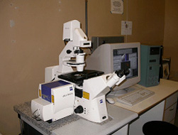
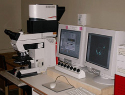
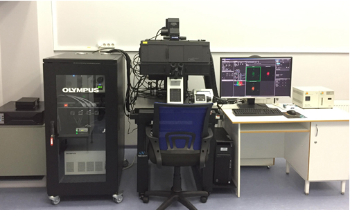
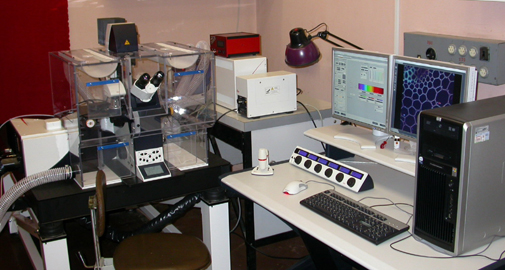
The Group was organized in 2002 to investigate scientific and methodological problems of confocal microscopy and image analysis, also to help researchers of Institute in their microscopic studies. Now the Group has laser scanning confocal microscopes OlLYMPUS FV3000, LEICA TCS SP5, fluorescent microscopes AXIOVERT 200M, AXIOPHOT and AXIOSKOP with digital videosystems. |
|
M.L. Vorobjev |
Scientific Staff:
S.V. Zherebtsov, Junior Research Scientist, Ph.D.
M.L. Vorobjev, Leading Engineer
|
Comparative features of laser scanning confocal microscopes
Selected Publications:
Bogolyubova I., Stein G., Bogolyubov D. FRET analysis of interactions between actin and exon-exon junction complex proteins in early mouse embryos.
Cell Tissue Res. 2013. 352: 277-285.
Agafonova N. A., Sakuta G.A., Rozanov Yu. M., Shtein G.I., Kudruavtsev B.N. DNA image-fluorimetry of individual human chromosomes. Cell and Tissue Biology.
2013. 7(4): 352-361.
Myasnikova E., Surkova S., Stein G., Pisarev A., Samsonova M. Regression system for estimation of errors introduced by confocal imaging into gene expression data
in situ. BMC Bioinformatics. 2011. 12: 20-331.
Bezborodkina N.N., Shtein G.I., Sivova E.V., Chestnova A.Yu., Kudryavtsev B.N. Analysis of structure of glycogen in rat hepatocytes using cytochemical and FRET methods.
Cell and Tissue Biology. 2011. 5(5): 417-427.
Egorova O.V., Shtein G.I. A comparision of fluorescence-microscope illuminator systems based on LEDs and HBO mercury lamps. J.Opt.Technol. 2011. 78(1): 81-83.
Stein G.I. Confocal microscopy and its application for analysis of cell cultures. In: Methods of cell cultivation (Eds. G. Pinaev, M. Bogdanova).
Publ. SPbSPU, 2008. p.261-274.
Stein G.I. Manual of confocal microscopy. Saint-Petersburg, Publ. SPbSPU, 2007, 77p.
Stein G.I.
Resolution of confocal microscope: evolution of ideas. 2006.
Stein G.I. Confocal microscopy: myths and reality. 2005.
Stein G.I. Laser scanning confocal microscopy. Manual. Publ. ICRAS. 2004. 32p.
Stein G.I. The influence of image analyser noises on the image quality in studies of cell texture features. Tsitologiya. 2002. 44 (1) : 102-106.
Microscope
Objectives
Temperature control
Laser lines (nm)
Main beamsplitter
Spectral confocal channels
Max frame resolution (pixel)
Scan zoom
Max line frequency (lin/s)
Resonant scanner (lin/s)
3D visualization
Spectral scanning
Colocalization
FRAP
FRET
Manufacturing
inverted
10×/0.4, 20×/0.7; 40×/1.25,
100×/1.4, 40×/0.55 (LD)
Вox
405, 458, 488, 514, 543, 633
AOBS
3 PMT (+BF/DIC)
8192×8192
1−64
2800
16 000
+
+
+
+
AB
2008
inverted
10×/0.3, 20×/0.5, 20×/0.45(LD),
40×/1.3, 40×/0.6 (LD), 60×/1.42
Box/stage
405, 488, 561, 640
dichroic mirrors
2 PMT+2 GaAsP (+BF/DIC)
4096x4096
1−50
2000
16 000
+
+
+
+
SE
2016
| Structure
| Home
|