| Laboratory of Cell Physiology |
Alexey A. VERENINOVPhD., DSc.phone: (812) 297-38-02
The Laboratory Staff:
|
|
|
The Corresponding Member of the USSR Academy of Sciences D.N. Nasonov simultaneously with the organization of the Institute of Cytology founded the Laboratory of Cell physiology. From 1957 to 1985 the Director of the Institute, Corresponding Member of the USSR Academy of Sciences, A.S. Troshin, headed the Laboratory. From 1986 until now the Laboratory is headed by A.A. Vereninov, PhD., DSc. The Laboratory had prepared several known scientists who later became heads of other laboratories and groups of the Institute: A.A. Lev (Group of physical chemistry of cell membranes, 1976), Academician N.N. Nikolsky, Director of Institute of Cytology in 1988-2004, Corresponding Member of the USSR Academy of Sciences G.N. Mozhaeva (Laboratory of ion channels of cell membranes, 1985), S.A. Krolenko, PhD., DSc. (Laboratory of biological cytology and cytochemistry, 1985). Pioneer trends in the cell study at the Laboratory were initiated by S.V. Levin, V.V. Malev, A.L. Krol (optical studies of cell motility underlying micromovement of nerve fiber during action potential and flickering erythrocytes and other non-excitable cells), A.B. Kaulin (study of anisotropic cell organelles by fluorescence polarization), and I.I. Marakhova (study of ion transport in proliferating cell cultures). |
|
After budding of daughter laboratories from the Laboratory of Cell physiology, two directions of work, both originating from the interests of D.N. Nasonov and A.S. Troshin, have been developed: (1) optical study of living cells and (2) study of ion balance in animal cells. The most remarkable results in the optical studies of living cells were the discovery of movement of nerve fiber during action potential and the finding that submicron local movements of cell surface within the range 0.2-30 Hz, which are observed in numerous non-muscle cells, were due not to the thermal or electrostriction fluctuations of lipid bilayer, but due to ATP-dependent motility of the membrane skeleton. The main publications related to optical studies of living cells: Levin S.V., Golfand K.A. 1980. Reversible displacement of crab axon at action potential. Tsitologiya. 22 (6) : 717-721 [in Russian]. Levin S.V., Golfand K.A.1984. Rapid transverse movement of crab axon at action potential. Tsitologiya. 26 (8) : 920-926 [in Russian]. Levin S.V., Golfand K.A., Malev V.V. 1986. Wave of local shortening and lengthening of single crab axon during action potential. Tsitologiya. 28 (12) : 1307-1315 [in Russian]. Levin S.V., Malev V.V. 1987. Effect of elastic properties of crab axon sheath on the nerve fiber movement during action potential. Tsitologiya. 29 (5) : 569-575 [in Russian]. Krol A.Yu., Grinfeldt M.G., Levin S.V., Smilgavichus A.D. 1990. Local mechanical oscillations of the cell surface within the range 0.2-30 Hz. Eur. Biophys. J. 19 : 93-99. Levin S., Korenstein R. 1991.Membrane fluctuations in erythrocytes are linked to MgATP-dependent dynamic assembly of the membrane skeleton. Biophys. J. 60 : 733-737. Krol A.Yu., Malev V.V., Grinfeldt M.G. 1993. Spectral characteristics of spontaneous oscillations in erythrocytes and their ghosts. Biol. Mem. 6 : 701-710. Rubashkin A., Iserovich P., Hernandez J.A., Fischbarg J. 2005. Epithelial fluid transport: protruding macromolecules and space charges can bring about electro-osmotic coupling at the tight junctions. J. Membrane Biol. 208 : 251-263. Tuvia S., Almagor A., Bitler A., Levin S., Korenstein R., Yedgar S. 1997. Cell membrane fluctuations are regulated by medium macroviscosity: evidence for a metabolic driving force. PNAS USA. 94 : 5045-5049. Tuvia S., Levin S., Bitler A., Korenstein R. 1998. Mechanical fluctuations of the membrane-skeleton are dependent on F-actin ATPase in human erythrocytes. J. Cell Biol. 141 : 1551-1561. Krol A.Yu., Malev V.V., Vereninov A.A. 2000. Laser dark-field microscope: investigation of the motility of submicroscopic structures. Proc. SPIE. 4064 : 45-48. Grinfeldt M.G., Krol A.Yu., Malev V.V. 2000. Correlation between the oscillatory and adhesion activities in erythroid cells. Membr. Cell Biol. 13 : 347-355. Krol A.Yu., Grinfeldt M.G., Vereninov A.A., Malev V.V. 2000. The role of actin cytoskeleton in the generation of surface oscillations of red blood cell ghosts. Membr Cell Biol. 14 (1) : 69-77. Studies of cell ion balance looked in the early 1960s at the two basic principles which might underlie the asymmetrical distribution of ions between the cytoplasm and the medium. These were, first, the principle of selective ion binding in cytoplasm as a phase, which was promoted from the 1930s by D.N. Nasonov and A.S. Troshin as an alternative to the classical membrane theory; and, second, the principle of perpetual ion pumping, which was advanced subsequently. 24Na, 22Na and 42K exchange in frog muscle were studied, and the data which had previously been considered as favoring the K+ binding in muscle were explained in terms of the pump-leak model. The results of this work were summarized in the book "Ion transport across the cell membrane. Analysis of fluxes" (1978) by A.A. Vereninov. All arguments for and against the "bulk phase" and "pump-leak" concepts were considered in this book; the possibility of determining individual activity coefficients of ions in the cytoplasm-phase was criticized; and a good deal of contemporary evidence was presented indicating that fluxes considered as "leaks" should be attributed to specific ion-transporting systems. Laboratory studies of ion transport in proliferating cell cultures began in the late 1970s. Significant changes were found in K+ and Na+ transport during the transition of resting cells to proliferation, indicating that high total K+ and Na+ content per g protein is a peculiar feature of all proliferating cells. At the same time, the idea that cell malignancy was associated with specific alteration of cell ion transport proved to be erroneous. The results of the first years of work on cell cultures were reported in the book: Vereninov A.A., Marakhova I.I (1986) "Ion transport in cultured cells". Further studies in the laboratory were developed along three lines, as follows: (1) The first line of studies involves detailed investigations of monovalent ion transport during G0-G1 transition, using activated human lymphocytes as a main model (Vereninov et al., 1991; Marakhova et al., 1995, 1998a,b, 1999). These studies include RT PCR assay of the expression of mRNAs coding for major ion transporters (АТР1В1, NHE1, NKCC1) in parallel with Bcl-2, p53, hSGK, GAPDH, and actin. The relation between common ("grouped") expression response and peculiarities in expression of individual mRNAs was investigated, as well as variability of the expression profiles in lymphocytes from different donors. It was shown that the node of the signaling network responsible for the grouped expression is located upstream of the elements responsible for individual variability (Vereninov et al., 2001a, b). Recent papers by Marakhova and colleagues (ref. see Karitskaya et al., 2010) make progress in further studying ion transport in activated lymphocytes at functional, protein and mRNA levels. (2) The second line of investigations concerns the changes in ion and water balance during apoptosis. An established experimental model of apoptosis in human lymphoid U937 cells and rat thymocytes treated with etoposide or staurosporine was used to measure the cellular Cl– content and fluxes, the K+, Na+ and water content, and ouabain-sensitive and ouabain-resistant Rb+ fluxes (Vereninov et al., 2003, 2004a, 2007, 2008; Yurinskaya et al 2005a, 2005b, 2010). Determination of caspase activity, confocal microscopy, and flow cytometry were used for testing apoptosis. It was found that no cell shrinkage took place in the case of apoptosis of U937 cells induced by etoposide, in contrast to apoptosis of the same cells induced by staurosporine, when shrinkage occurs. These results show that the classical definition of apoptosis as a "shrinkage-necrosis" is inadequate. Changes in water, K+, Na+, Cl– content, as well as changes in the content of other intracellular osmolytes during apoptosis, were estimated quantitatively for the first time. Rearrangement of the monovalent ion fluxes underlying these changes was also revealed using Rb+, 22Na+ and 36Cl– tracers (Vereninov et al., 2007, 2008; Rubashkin et al., 2010). (3) A mathematical model of the overall unidirectional K+, Na+, and Cl– flux balance in normal and apoptotic cells was developed (Vereninov, Vereninov, 1991; Vereninov et al., 1995, 1997, 2004b, 2006, 2007; Rubashkin et al., 2010). Systemic application of this model to the experimentally measured fluxes facilitated a comprehensive description of changes in monovalent cation fluxes and water balance during apoptosis. This description could not have arisen from electrophysiological, radiotracer, or inhibitor studies alone. In particular, a decrease in the permeability of the Na+ channels proved to be crucial in preventing cell swelling due to the decrease in Na+/K+ pump activity in cells undergoing apoptosis. It was further found that apoptotic cell shrinkage may occur without extra-opening of the K+ and Cl– channels. In view of this finding, and abundant electrophysiological evidence that K+ and Cl– channels are activated during apoptosis, it has been proposed that the activity of K+ and Cl– channels during apoptosis is relevant to the control of the plasma membrane potential - which may in turn have a regulatory function in apoptosis. Electro-osmosis as a driving force in paracellular fluid transport in epithelia is studied in the laboratory by A.A. Rubashkin. The role of the fixed electric charge in tight junctions, e.g. associated with claudine has been studied theoretically and examined in experiments with corneal endothelium due to collaboration with colleagues from Columbia University (Sanchez et al., 2002; Rubashkin et al., 2005; Rubashkin 2006; Fischbarg et al., 2006). Their paper (Rubashkin et al., 2005) was selected by "Faculty of 1000 Biology" as one of the most interesting 1000 recent studies in biology).
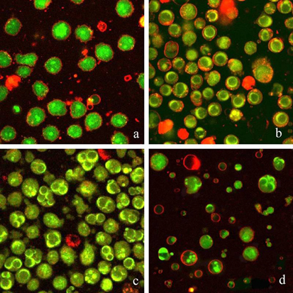
Apoptosis of U937 cells Confocal images of human lymphoid cells U937 stained with acridine orange, ethidium bromide, and the lipophilic dye RH414. a - control cells, b, c - apoptosis caused by staurosporine (b) or etoposide (c), d - apoptotic bodies (Yurinskaya et al. Cell Physiol. Biochem. 2005. 16 : 155-162). The main publications related to studies of ion balance in animal cells since 1991: Vereninov A.A., Vereninov A.A.,Jr. 1991. Ionic, electrical and water balance in animal cell. A model with the Na,K-ATPase active transport, Goldman channels and Na+K+2Cl symporter. Tsitologiya. 33 (11) : 4-17 [in Russian]. Vereninov A.A., Gusev T.V., Kazakova O.M., Klimenko E.M., Marakhova I.I., Osipov V.V., Toropova F.V. 1991. Transport and distribution of monovalent cations in human peripheral blood lymphocytes activated by phytohemagglutinin. Tsitologiya. 33 (11) : 78-93 [in Russian] . Marakhova I.I., Vereninov A.A., Toropova F.V., Vinogradova T.A. 1995. Long-term enhancement of Na, K-ATPase pump during blasttransformation of human lymphocytes is controlled first by translational, then by transcriptional mechanisms. FEBS Lett. 368 : 110-112. Vereninov A.A., Glushankova L.N., Rubashkin A.A. 1995. The role of ion transport in long-term cell volume regulation. Mathematical model and live lymphoid cells. Tsitologiya. 37 (12) : 1151-1166 [in Russian] . Vereninov A.A., Glushankova L.N., Rubashkin A.A. 1997. Effect of NaK2Cl symport and chloride channel permeability on the ion flux balance and ion distribution in animal cells of different types. Tsitologiya. 39 (8) : 727-739. Marakhova I.I., Vereninov A.A., Toropova F.V., Vinogradova T.A. 1998. Na, K-ATPase pump in activated human lymphocytes: on the mechanisms of rapid and long-term increase in K influxes during blasttransformation. Biochim. Biophys. Acta. 1368 : 61-72. Marakhova I.I., Vereninov A.A., Vinogradova T.A. Toropova F.V. 1998. Cyclosporin A inhibits long-term activation of Na,K pump in phytohemagglutinin-stimulated human lymphocytes. Membr. Cell Biol. 12 : 363-374. Marakhova I.I., Ivanova A.E., Toropova F.V., Vereninov A.A., Vinogradova T.A. 1999. Functional expression of the Na/K pump is controlled via a cyclosporin A-sensitive signalling pathway in activated human lymphocytes. FEBS Letters. 456 : 285-289. Vereninov A.A., Vasilieva I.O., Yurinskaya V.E., Matveev V.V., Glushankova L.N. 2001a. A study of the "group" expression of ATP1B1, NHE1, NKCC1, β-actin, GAPDH, p53, BCl-2, and hSGK mRNAs in human lymphocytes activated with phytohemagglutinin. Tsitologiya. 43 (6) : 602-612 [in Russian] . Fillon S., Warntges S., Matskevitch J., Moschen I., Setiawan I., Gamper N., Feng Y.X., Stegen C., Friedrich B., Waldegger S., Broer S., Wagner C.A., Huber S.M., Klingel K., Vereninov A., Lang F. 2001. Serum- and glucocorticoid-dependent kinase, cell volume, and the regulation of epithelial transport. Comp. Biochem. Physiol., Part A. 130 : 367-376. Vereninov A.A., Vassilieva I.O., Yurinskaya V.E., Matveev V.V., Glushankova L.N., Lang, F., Matskevich J.A. 2001b. Differential transcription of ion transporters, NHE1, ATP1B1, NKCC1 in human peripheral blood lymphocytes activated to proliferation. Cell. Physiol. Biochem. 11 (1) : 19-26. Zuniga F.A., Shi G., Haller J.F., Rubashkin A., Flynn D.R., Iserovich P., Fischbarg J. 2001. A three-dimensional model of the human facilitative glucose transporter Glut1. J. Biol. Chem. 276 : 44970-44975. Sanchez J.M., Li Y., Rubashkin A., Iserovich P., Wen Q., Ruberti J.W., Smith R.W., Rittenband D., Kuang K., Diecke F.P.J., Fischbarg J. 2002. Evidence for a central role for electro-osmosis in fluid transport by corneal endothelium. J. Membrane Biol. 187 : 37-50. Vereninov A.A., Volgareva E.V., Matveev V.V., MoshkovA.V., Rozanov Yu.M., Shirokova A.V., Yurinskaya V.E. 2003. Water and ion balance of rat thymocytes in apoptosis induced by dexamethasone or etoposide. Ion-osmotic model of cell volume decrease. Tsitologiya. 45 (5) : 500-509 [in Russian]. Vereninov A.A., Goryachaya T.S., Matveev V.V., Moshkov A.V., Rozanov Yu.M., Sakuta G.A., Shirokova A.V., Yurinskaya V.E. 2004a. Cell shrinkage in apoptosis is not obligatory. Apoptosis of U937 cells induced by staurosporine and etoposide. Tsitologiya. 46 (7) : 609-619 [in Russian]. Vereninov A.A., Yurinskaya V.E., Rubashkin A.A. 2004b. The role of potassium, potassium channels, and symporters in apoptotic cell volume decrease: Experiment and theory. Doklady Biological Sciences, 398 : 417-420. Yurinskaya V.E., Moshkov A.V., Rozanov Yu.M., Shirokova A.V., Shumilina E.V., Vassilieva I.O., Lang F., Volgareva E. V, Vereninov A.A. 2005a. Thymocyte K+, Na+ and water balance during dexamethasone and etoposide induced apoptosis. Cell. Physiol. Biochem. 16 : 15-22. Yurinskaya V.E., Goryachaya T.S., Moshkov A.V., Rozanov Yu.M., Sakuta G.A, Shirokova A.V., Shumilina E.V., Vassilieva I.O., Lang F., Vereninov A.A. 2005b. Potassium and sodium balance in U937 cells during apoptosis with and without cell shrinkage. Cell. Physiol. Biochem. 16 : 155-162. Lang F., Foller M., Lang K., Lang P., Wieder T., Ritter M., Gulbins E., Vereninov A., Huber S. 2005. Ion channels in cell proliferation and apoptotic cell death. J. Membr. Biol. 205 (3) : 147-157. Rubashkin A., Iserovich P., Hernandez J., Fischbarg J. 2005. Epithelial fluid transport: protruding macromolecules and space charges can bring about electro-osmotic coupling at the tight junctions. J. Membr. Biol. 208 : 251-263. Fischbarg J., Diecke F.P.J., Iserovich P., Rubashkin A. 2006. The role of the tight junction in paracellular fluid transport across corneal endothelium. Electro-osmosis as a driving force. J. Membr. Biol. 210 (2) : 117-130. Rubashkin A.A. 2006. A model of electro-osmosis in a leaky tight junction of epithelial cells. Doklady Biochemistry and Biophysics. 407 : 71-73. Lang F., Shumilina E., Ritter M., Gulbins E., Vereninov A., Huber S.M. 2006. Ion channels and cell volume in regulation of cell proliferation and apoptotic cell death. Contrib. Nephrol. 152 : 142-160. Vereninov A.A., Yurinskaya V.E., Rubashkin A.A. 2006. Apoptotic shrinkage of lymphoid cells: a model of changes in ion flux balance. Doklady Biochemistry and Biophysics. 411 : 356-360. Vereninov A.A., Goryachaya T.S., MoshkovA.V., Vassilieva I.O., Yurinskaya V.E., Lang F., Rubashkin A.A. 2007. Analysis of the monovalent ion fluxes in U937 cells under the balanced ion distribution: Recognition of ion transporters responsible for changes in cell ion and water balance during apoptosis. Cell Biol. Intern. 31 (4) : 382-393. Shirokova A.V. 2007. Apoptosis signaling pathways and cell ion and water balance. Cell and Tissue Biology. 1 (3) : 215-224. Rubashkin A.A., Iserovich P. 2007. A new approach to the selectivity of ion channels: nonlocal electrostatic consideration. Dokl. Biophys. Biochem. 417 : 302-305. Lang F., Foller M., Lang K., Lang P., Ritter M., Vereninov A., Szabo I., Huber S.M., Gulbins E. 2007. Cell volume regulatory ion channels in cell proliferation and cell death. Methods in Enzymology. Osmosensing and Osmosignaling. 428 : 209-225. Lang F., Gulbins E., Szabo I., Vereninov A.A., Huber S.M. 2008. Ion channels, cell volume, cell proliferation and apoptotic cell death. In: Sensing with Ion Channels. Springer Series in Biophysics 11. B.Martinac (ed.). Vereninov A.A., Rubashkin A.A., Goryachaya T.S., Moshkov A. V., Rozanov Y.M., Shirokova A.V., Strelkova E.G., Lang F., Yurinskaya V.E. 2008. Pump and channel K (Rb+) fluxes in apoptosis of human lymphoid cell line U937. Cell Physiol. Biochem. 22 : 187-194. Rubashkin A.A., Yurinskaya V.E., Vereninov A.A. 2010. . Calculations of K+, Na+ and Cl– fluxes across cell membrane with Na+/K+ pump, NKCC, NC cotransport and ionic channels with non-Goldman rectification in K+-channels: normal and apoptotic cells. Cell and Tissue Biology. 4 (5) : 464-470. Yurinskaya V.E., Goryachaya T.S., Rubashkin A.A., Shirokova A.V., Vereninov A.A. 2010. Changes in K+, Na+ and Cl– balance and K+ and Cl– fluxes in U937 cells induced to apoptosis by staurosporine: on cell dehydration in apoptosis. Cell and Tissue Biology. 4 (5) : 457-463. Yurinskaya V.E., Rubashkin A.A., Shirokova A.V., Vereninov A.A. 2011. Regulatory Volume Increase (RVI) and Apoptotic Decrease (AVD) in U937 Cells in Hypertonic Medium. Сell and Tissue Biology, 2011. Vol. 5. P. 487-494. Yurinskaya V. E., Rubashkin A. A., Vereninov A. A. 2011. Balance of unidirectional monovalent ion fluxes in cells undergoing apoptosis: why does Na+/K+ pump suppression not cause cell swelling? Journal of Physiology, Vol. 589, N. 9. P. 2197-2211. Yurinskaya V. E., Moshkov A. V., Wibberley A. V., Lang F., Model M. A., Vereninov A. A. 2012. Dual response of human leukemia U937 cells to hypertonic shrinkage: initial regulatory volume increase (RVI) and delayed apoptotic volume decrease (AVD). Cell. Physiol. Biochem. Vol. 30. P. 964-973. Vereninov I.A., Yurinskaya V.E., Vereninov A.A. 2013. Integration of monovalent ion fluxes across plasma membrane under the balanced state: ion gradients and water balance in animal cells. Russian Journal of Physiology. 99 (5) ; 619-629. Vereninov I.A, Vereninov A.A. 2013. Algoritmy komp'iuternogo modelirovaniia perenosa ionov v kletkakh [Algorithms of computer modelling of ion interaction in cells] (rus). St Petersburg State Polytechnical University Journal Computer Science Telecommunication and Control System 169: 91-97. Yurinskaya V.E., Moshkov A.V, Goryachaya T.S, Vereninov A.A. 2014. Li/Na exchange and Li active transport in human lymphoid cells U937 cultured in lithium media. Cell and Tissue Biology 8: 80-90. Vereninov I.А., Yurinskaya V.E., Model М.А., Lang F., Vereninov A.A. 2014. Computation of Pump-Leak Flux Balance in Animal Cells. Cell Physiol. Biochem. Vol. 34:1812-1823. DOI: 10.1159/000366382 Vereninov I.A., Vereninov A.A., Yurinskaya V.E. 2014. Supplement for computation of monovalent ion flux balance in animal cells:
|